National Cytotechnology Day
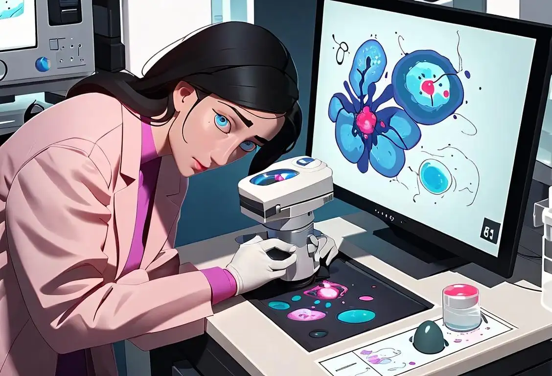
Hey there cytotechnology enthusiasts! Get ready to celebrate National Cytotechnology Day, a day dedicated to recognizing the important work of cytotechnologists. So grab your microscopes and let's dive into the fascinating world of cytotechnology!
When is Cytotechnology Day?
It's national cytotechnology day on the 13th May.
The Wonders of Cytotechnology
Have you ever wondered how doctors diagnose diseases like cancer? Well, that's where cytotechnologists come to the rescue! They are the unsung heroes of the medical field who specialize in examining cells under a microscope to detect abnormalities.
On National Cytotechnology Day, we celebrate these talented professionals who play a crucial role in early disease detection. They meticulously examine cell samples, identify cancerous or pre-cancerous cells, and help medical professionals make accurate diagnoses.
Cytotechnologists are like the detectives of the cell world. Armed with their trusty microscopes, they carefully analyze cellular clues and provide vital information to help save lives. So, let's raise a glass (preferably a microscope slide) to these unsung heroes!
The Internet and National Cytotechnology Day
While National Cytotechnology Day may not have a long history on the internet, it's gaining recognition and appreciation year after year. Social media platforms, medical websites, and online communities come alive on May 13th to honor cytotechnologists and spread awareness about their invaluable contributions.
Whether it's sharing stories of survival, thanking cytotechnologists, or highlighting groundbreaking research in the field, the internet plays a vital role in bringing people together to celebrate this special day. So, let's virtually gather around and show our support for these amazing professionals online!
History behind the term 'Cytotechnology'
1950
Emergence of Cytotechnology
In the year 1950, the term 'cytotechnology' emerged as a pioneering field in the medical sciences. Stemming from the Greek word 'kytos' meaning 'cell,' cytotechnology refers to the study of cells, particularly under a microscope. It encompasses the examination and analysis of the cellular structure, function, and abnormalities, aiding in the early detection and diagnosis of various diseases.
1934
The Birth of Cytotechnology
Cytotechnology, the study and examination of cells for diagnostic purposes, finds its origins in 1934. That year, a pathologist named George Papanicolaou discovered that abnormalities in cervical cells could be used to detect early signs of cervical cancer. This groundbreaking finding laid the foundation for cytotechnology as a distinct field within diagnostic medicine.
1969
The Birth of Cytotechnology
Cytotechnology was first recognized as a unique field of study in 1969. It involves the examination of cells for the identification and classification of diseases. With the advancement of technology and the need for specialized professionals, cytotechnology emerged as a valuable discipline in the medical field.
1952
First Use of the Term
The term 'cytotechnology' was first used in the year 1952. It was coined by Dr. George Papanicolaou, a Greek-American physician and pioneer in the field of cytopathology. He used the term to describe the science and practice of examining cells to diagnose diseases.
1928
The Discovery of Pap Smear
In 1928, Dr. George Papanicolaou introduced the revolutionary Pap smear test. Initially, this test was used to detect early signs of cervical cancer by examining cells from the cervix. The Pap smear quickly became an effective tool in diagnosing various gynecological conditions.
1950
The Birth of Cytotechnology
Cytotechnology is a term that was coined in 1950. It originates from the fusion of two words: 'cyto', which means cell, and 'technology', which refers to the application of scientific knowledge for practical purposes. Cytotechnology, therefore, refers to the field of studying cells through various laboratory techniques.
1969
Emergence of cytotechnology
Cytotechnology, a term coined by George N. Papanicolaou, came into existence in 1969. It encompasses the study and interpretation of cells for the detection of cancer and other diseases. Papanicolaou, also known as the 'Father of Cytology,' revolutionized the field with his development of the Pap smear, a screening test for cervical cancer. This marked the beginning of a new era in the field of healthcare.
1946
The Birth of Cytotechnology
In 1946, the term 'cytotechnology' was first coined by George N. Papanicolaou, a Greek-American physician and researcher. Papanicolaou developed a technique for examining cells under a microscope, known as the Papanicolaou stain or Pap smear. This breakthrough allowed for the early detection of cervical and uterine cancer through the examination of cellular abnormalities. With the introduction of this technique, cytotechnology, the study and analysis of cells, became a crucial field in the realm of medicine.
1969
Formation of the Cytotechnology Board
During the year 1969, in response to the increasing importance of cytotechnology as a discipline, the American Society of Clinical Pathologists (ASCP) established the Cytotechnology Board. This board aimed to ensure the maintenance of high professional standards and competence among cytotechnologists by developing certification programs and conducting examinations. It played a pivotal role in shaping the education and practice of cytotechnology.
1957
The Cytotechnologist Profession
In 1957, the cytotechnologist profession was officially recognized. The American Society of Cytology, now known as the American Society of Cytopathology, established guidelines for the training and certification of cytotechnologists. Cytotechnologists are highly trained professionals who specialize in the microscopic examination of cells to identify early signs of cancer or other abnormalities. They work closely with pathologists to provide accurate and timely diagnoses, playing a vital role in the early detection and prevention of various diseases.
1956
The Introduction of the Papanicolaou Test
In 1956, George Papanicolaou introduced the Papanicolaou test, commonly known as the Pap smear. This screening method became a crucial tool in cytotechnology as it allows the detection of abnormal cells in cervical samples. The Pap smear has since saved countless lives by enabling early detection of cervical cancer.
1971
Cytotechnology Accreditation
In 1971, the Commission on Accreditation of Allied Health Education Programs (CAAHEP) approved the first accreditation standard for cytotechnology programs. This step ensured that the education and training of cytotechnologists met specific criteria, leading to a higher quality of healthcare and diagnostics for patients. Accreditation also provided recognition and legitimacy to the field.
1950
Pap Smears Introduce Cytotechnologists
In the 1950s, the Pap smear test gained popularity as a way to screen for cervical cancer. This test involved collecting cervical cell samples, staining and preparing them on slides, and examining them under a microscope. With this increased demand for analyzing cell samples, the need for specialists skilled in cytology arose. Cytotechnologists became the specialists responsible for evaluating these specimens, identifying abnormalities, and helping with early cancer detection.
1948
Cytotechnology as a Profession
In 1948, cytotechnology emerged as an independent profession. Cytotechnologists are trained specialists who analyze cells and cellular abnormalities to help diagnose diseases. They play a crucial role in the early detection and prevention of various cancers and other medical conditions.
1961
Creation of the Cytotechnology Profession
In 1961, the American Society of Cytology recognized cytotechnology as a distinct profession. This recognition marked an important milestone in the field, as it highlighted the significance of cytotechnologists in the early detection and diagnosis of various diseases, especially cancer.
1971
The birth of cytotechnology education programs
In 1971, the first cytotechnology education programs were established in the United States. These programs aimed to train professionals in the specialized techniques and skills required for cytological analysis. With the rise in demand for accurate and timely diagnoses, cytotechnology education programs played a crucial role in producing competent cytotechnologists.
1969
The Formation of the American Society of Cytotechnology
1969 marked an important milestone in the development of cytotechnology. The American Society of Cytotechnology (ASCT) was established to promote and advance the profession. The ASCT has been instrumental in setting standards for education, certification, and professionalism in the field, ensuring that cytotechnologists provide accurate and reliable results.
1983
The Papanicolaou Society of Cytopathology
The Papanicolaou Society of Cytopathology (PSC) was established in 1983. Named after Dr. George Papanicolaou, the inventor of the Pap smear, the society aimed to promote scientific advancements and education in cytology. The PSC plays a crucial role in enhancing the knowledge and skills of cytotechnologists through conferences, publications, and collaborations.
1970
Professional Recognition and Accreditation
Cytotechnology started gaining recognition as a distinct profession in the 1970s. Professional organizations such as the American Society of Cytopathology (ASC) and the American Society of Clinical Pathologists (ASCP) were established to promote high standards and provide resources for cytotechnologists. Accreditation programs for cytotechnology schools were also introduced, ensuring that professionals in this field received proper training and education.
1983
Recognition by the National Accrediting Agency for Clinical Laboratory Sciences
The year 1983 witnessed a significant milestone for cytotechnology with its recognition as a profession by the National Accrediting Agency for Clinical Laboratory Sciences (NAACLS). This recognition brought standardization and quality assurance to the field, ensuring that cytotechnologists meet the required educational and competency standards.
1970
Inclusion in the Clinical Laboratory Improvement Amendments (CLIA)
In 1970, cytotechnology was included in the Clinical Laboratory Improvement Amendments (CLIA), which are regulatory standards for laboratory testing. This recognition solidified the importance of cytotechnologists' contributions to healthcare, ensuring proper training, and quality control in cytology laboratories.
1980
Advancements in Cytotechnology
In 1980, technological advancements greatly enhanced the field of cytotechnology. Automated screening systems and digital imaging revolutionized the way cytotechnologists analyze cells. These advancements allowed for faster and more accurate diagnoses, reducing the need for manual screening of slides. Cytotechnologists began utilizing computer-assisted screening tools to assist in the detection of abnormal cells, improving the efficiency and precision of their work.
1978
National Recognition for Cytotechnology
In 1978, the U.S. Congress acknowledged the significance of cytotechnology as an important field in healthcare by passing the Clinical Laboratories Improvement Amendments (CLIA). These amendments recognized cytotechnologists as an integral part of the laboratory team and established specific quality standards for cytology laboratories. This national recognition further solidified the role of cytotechnology in the medical community.
1973
Formation of Accredited Cytotechnology Programs
In 1973, the first accredited cytotechnology programs were established in the United States. These programs provided comprehensive training and education to aspiring cytotechnologists, ensuring a standardized level of competence and knowledge in the field.
1998
The Cytotechnology Recognition Letter
In 1998, the Centers for Medicare and Medicaid Services (CMS) implemented the Cytotechnology Recognition Letter (CRL) program. This program evaluated and recognized cytotechnologists who demonstrated exceptional proficiency in screening and interpreting gynecological and nongynecological cell specimens. The CRL program aimed to ensure a high standard of quality in cytotechnologist performance, contributing to the continued improvement of cytotechnology as a vital healthcare discipline.
1991
Recognition by the American Society of Clinical Pathologists
The American Society of Clinical Pathologists (ASCP) formally recognized cytotechnology as a distinct laboratory science profession in 1991. This recognition further solidified the importance of cytotechnologists in the field of pathology and their contributions to patient care.
1983
Accreditation of Cytotechnology Programs
The year 1983 marked a significant milestone as the Commission on Accreditation of Allied Health Education Programs (CAAHEP) granted accreditation to cytotechnology programs. This accreditation ensured that the educational institutions offering cytotechnology courses met established standards, thus ensuring the quality of training received by aspiring cytotechnologists.
1990
Advancements in Technology and Automation
The 1990s brought significant advancements in technology, which revolutionized cytotechnology. Automated screening systems and computer-assisted analysis tools improved the efficiency and accuracy of evaluating cell samples. These technological advancements allowed cytotechnologists to process larger volumes of specimens and detect abnormalities with greater precision.
1992
National Cytotechnology Day
In 1992, the American Society of Cytopathology (ASC) declared May 13th as National Cytotechnology Day. This day celebrates the contributions of cytotechnologists to patient care and recognizes their expertise in identifying cellular abnormalities. National Cytotechnology Day serves as a reminder of the critical role played by cytotechnologists in early disease detection and prevention.
1995
Advancements in Automation and Digital Imaging
The field of cytotechnology witnessed significant advancements in automation and digital imaging technology in 1995. This revolutionized the way cells were analyzed and allowed for more efficient and accurate detection of abnormal cellular changes. Digital imaging also facilitated collaboration among cytotechnologists and pathologists.
1990
Advancements in Immunocytochemistry
In the 1990s, significant advancements in immunocytochemistry expanded the capabilities of cytotechnology. This technique utilizes specific antibodies to identify and analyze proteins within cells. Immunocytochemistry has become a valuable tool for diagnosing various diseases and identifying specific cell types, enhancing the accuracy and efficiency of cytotechnologists' work.
2004
Cytotechnology Week
For raising awareness and recognizing the contributions of cytotechnologists, the American Society of Cytopathology designated the first full week of May as Cytotechnology Week in 2004. This annual celebration honors the skill, expertise, and dedication of cytotechnologists who play a vital role in the early detection and prevention of various diseases.
2008
Transition to Digital Imaging
Around 2008, the field of cytotechnology began transitioning from traditional microscopy to digital imaging. Digital imaging systems allow cytotechnologists to capture high-resolution images of cells, facilitating remote interpretation, sharing, and consultation. This technological shift has improved efficiency, collaboration, and documentation within the field.
2007
Cytotechnology Evolves with Molecular Diagnostics
As the field of molecular diagnostics expanded, cytotechnology adapted to incorporate new techniques. Molecular cytotechnology emerged, integrating molecular testing methods to analyze cells at a genetic level. This evolution enabled cytotechnologists to provide more detailed information on cellular changes, leading to enhanced diagnostic capabilities and personalized treatment approaches.
1994
Introduction of Liquid-based Cytology
1994 witnessed a major advancement in cytotechnology with the introduction of liquid-based cytology (LBC). This technique revolutionized the process of preparing and examining cells by replacing the traditional slide-based method. LBC offered improved clarity, reduced sampling errors, and enhanced the accuracy of diagnoses, significantly impacting the field of cytotechnology.
2004
The Evolution of Cytotechnology
In 2004, the field of cytotechnology continued to evolve as new techniques and technologies were developed. Molecular cytopathology emerged, combining traditional cytology methods with DNA and RNA analysis. This integration allowed for more comprehensive cellular analyses, aiding in the accurate diagnosis and treatment of various diseases. Additionally, cytotechnologists started exploring telecytology, a practice that enables remote viewing and diagnosis of cell specimens, improving access to pathology expertise in underserved areas.
2013
Integrating Molecular Techniques
In 2013, cytotechnology further evolved with the integration of molecular techniques into routine testing. This inclusion enabled cytotechnologists to detect and analyze genetic and molecular alterations in cells, improving the accuracy of diagnoses and tailoring treatment strategies.
2005
Advancements in Digital Imaging
With the advancements in digital imaging technology, cytotechnology underwent a significant transformation in 2005. Digital imaging allowed for more efficient and accurate analysis of cellular specimens, aiding in the early detection and diagnosis of diseases.
2004
Digital Cytology
With the advancement of technology, digital cytology emerged as a significant development in the field. In 2004, the American Society of Cytopathology organized the first conference on digital cytology to explore the potential of digital imaging and telecytology. Digital cytology revolutionized the way cytotechnologists analyze and share cell images, enabling faster and more accurate diagnoses.
Present
Advancements in digital cytotechnology
Today, cytotechnology continues to evolve with advancements in digital imaging and telecytology. Digital cytotechnology enables the interpretation and analysis of cell samples using digital images, improving accuracy and efficiency. Telecytology allows remote sharing and consultation of cytology cases, facilitating collaboration and access to expertise regardless of geographical barriers. These technological advancements have further enhanced the field of cytotechnology.
2019
Evolution of Molecular Cytotechnology
In recent years, the field of cytotechnology has expanded to incorporate molecular techniques. Molecular cytotechnology involves the study of cellular structures and functions at a molecular level. This development has further enhanced the diagnostic capabilities of cytotechnologists and opened up new avenues for research and treatment.
2009
Recognition of Molecular Cytotechnology
In the year 2009, the term 'molecular cytotechnology' emerged in response to the rapid advancement of molecular biology techniques. Molecular cytotechnology involves the analysis of cells at a molecular level, integrating molecular biology and cytology to improve the understanding and diagnosis of diseases such as cancer. This recognition further expanded the scope of cytotechnology in the era of molecular medicine.
Did you know?
Did you know that cytotechnologists can examine up to 100,000 cells per day? Talk about impressive multitasking skills!Tagged
awareness science healthcareFirst identified
13th May 2015Most mentioned on
13th May 2020Total mentions
46Other days

Genetic Counselor Awareness Day
Cytotechnology Day

Periodic Table Day

Stem Day

Fossil Day

Physician Suicide Awareness Day

Period Day

Nurses Day

Healthcare Decisions Day

Dna Day